Style Sampler
Layout Style
Patterns for Boxed Mode


Backgrounds for Boxed Mode
Search News Posts
General Inquiries 1-888-555-5555
•
Support 1-888-555-5555

Medica Image Analysis
Medical images represent the procedures used to image the inside(interior) of a patient’s body for clinical analysis. It is generated commonly by using X-rays, Magnetic Resonance Imaging (MRI), Microscopy Image, Optical Coherence Tomography (OCT), or Position Emission Tomography (PET)
Clinical data comes not only in the form of clinical notes or images, but also in many other forms like demographic notes (Gender, age, location, and marital status), results of laboratories tests, physiological measurement results, etc.
Medical image segmentation is a crucial task in medical imaging and plays a significant role in diagnosis, treatment planning, and monitoring of various diseases. It involves identifying and delineating regions of interest within medical images such as X-rays, CT scans, MRIs, and ultrasound images. Segmentation helps in the accurate localization of structures and abnormalities in the medical images, which can aid in better diagnosis and treatment planning.
The segmentation process involves dividing an image into multiple regions that correspond to different anatomical structures or pathological lesions. This task is challenging because of the complex nature of medical images, which may contain noise, artifacts, and low contrast between different tissues. Therefore, it is essential to use advanced image processing techniques and algorithms for accurate and reliable segmentation.
Various techniques have been proposed for medical image segmentation, ranging from manual segmentation to automated segmentation using machine learning algorithms. Manual segmentation involves a human expert drawing the region of interest on the image using a digital tool. While manual segmentation is accurate, it is time-consuming, and inter-observer variability can affect the results.
In contrast, automated segmentation algorithms use computational methods to identify regions of interest within medical images. These methods can be broadly classified into two categories: classical segmentation techniques and deep learning-based methods.
Classical segmentation techniques include thresholding, region-growing, edge detection, and active contour models. These techniques use mathematical operations to identify regions of interest within an image. While classical techniques can be fast and efficient, they often require fine-tuning of parameters for each image, which can be time-consuming and require expert knowledge.
Deep learning-based methods have revolutionized medical image segmentation in recent years. These methods use convolutional neural networks (CNNs) to learn features directly from the images and generate segmentation maps. CNNs have shown remarkable performance in various medical image segmentation tasks, including brain tumor segmentation, lung nodule detection, and liver segmentation.
One of the key advantages of deep learning-based methods is their ability to learn from large datasets, which can improve the accuracy and generalizability of the segmentation results. Moreover, these methods can handle complex and noisy images, making them suitable for various medical imaging applications.
Despite the remarkable performance of deep learning-based methods, they also have some limitations. These methods require a large amount of annotated data for training, which can be time-consuming and expensive to acquire. Additionally, the interpretability of these models is limited, and the black-box nature of the models can make it challenging to understand how they arrived at their segmentation results.
In conclusion, medical image segmentation is a critical task in medical imaging, with numerous applications in diagnosis, treatment planning, and monitoring of various diseases. Classical segmentation techniques and deep learning-based methods have their strengths and weaknesses, and the choice of method depends on the specific application and available resources. With the rapid advancement in computer vision and machine learning techniques, medical image segmentation is expected to continue improving and help healthcare professionals make more accurate and informed decisions.
Medical image segmentation is the task of segmenting objects of interest in a medical image. The field of biomedical imaging has exploded in recent years and anaplatform uses latest technologies in the field. We can navigate through whole-body CT scan, segment a cardiac MRI time series, and determine whether Alzheimer’s disease changes brain structure.
anaplatform makes it easy to add image and video analytics to your applications. You only provide an image or video to the MainPlatform, and the service can identify objects, people, text, scenes, and events. anaplatform has a simple, easy-to-use API that can quickly analyze any image or video.
Medical images are an integral part of the patient's electronic health records and they play an important role in helping patients diagnose and treat patients in health organizations
Medical images, ultrasound, X-ray (X-ray), CT, MR, Positron Emission Tomography briefly PET (Positron Emission Tomography), retinal photography and dermoscopy imaging consists of images obtained by the methods. imaging such as CT and MRI retinal photography and dermoscopy methods examine multiple organs. is specific.
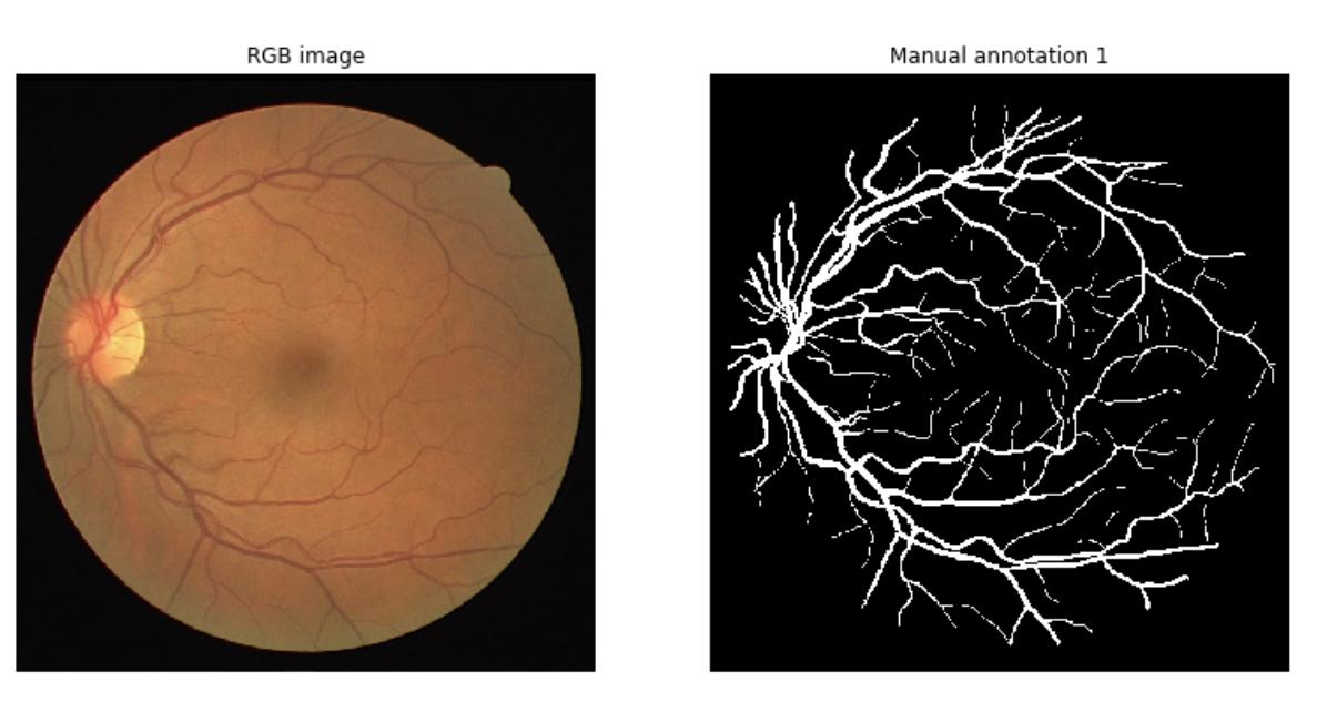
The exctraction of retinal vasculature from eye fundus images is a fundamental task in retinal image analysis. Over recent years, increasingly complex approaches based on sophisticated Convolutional Neural Network architectures have been pushing performance on well-established benchmark datasets.
Retinal vessel segmentation is one of the first and most important tasks for the computational analysis of eye fundus images. It represents a stepping stone for more advanced applications such as artery/vein ratio evaluation11, blood flow analysis5, image quality assessment12, retinal image registration13 and synthesis.
Our services has proven that there is no need of designing complex CNN architectures to outperform most current techniques on the task of retinal vessel segmentation, and (2) when a state-of-the-art model is trained on a particular dataset and tested on images from different data sources.
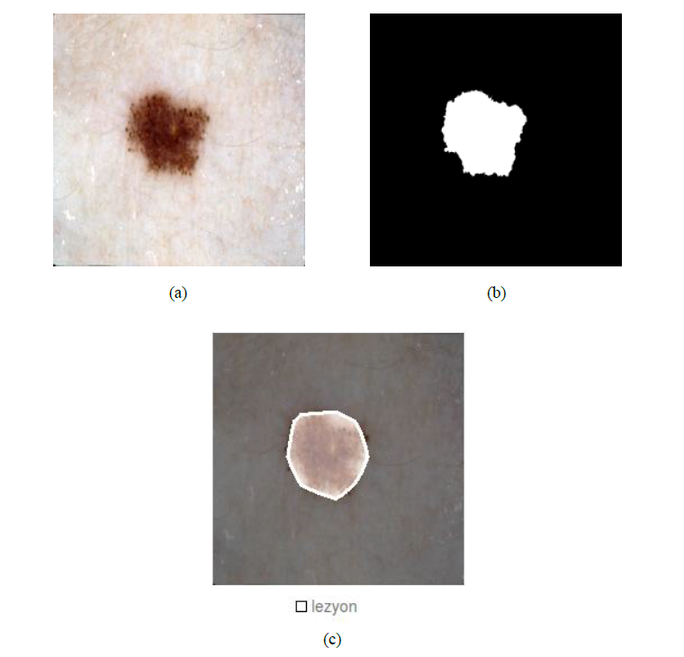
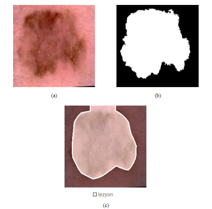
Skin lesion segmentation is performed to assist in the detection of melanoma, the most dangerous type of skin cancer.
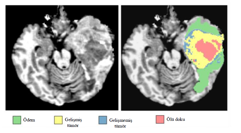
Each pixel in a group is a whole, as shown on the medical image below. corresponds to the object class. In a medical image important to the diagnosis of the disease these classes; There may be a background with a lesion or tumor. Label images of medical images, It specifies the pixel position of the anatomical feature that the deep neural network wants to learn.
An example test image with FCN-AlexNet trained using the pre-trained model for lesion segmentation-1: (a) Test image, (b) Definitive reference image, (c) Segmentation inference
With a training data from more than 400 open-access MR images, we can do registration, resampling, and image comparison and use the extracted measurements to evaluate the effect of Alzheimer's Disease on brain structure
With our Image Analysis service, you can apply different filters to images and obtain a histogram map. The anaplatform image service provides a comprehensive set of reference standard algorithms and workflow applications for analysis, visualization, and algorithm development. You can perform image segmentation, image enhancement, noise reduction, geometric transformations and image recording using deep learning and traditional image processing techniques. Toolbox supports processing of 2D, 3D and arbitrarily large images.
Analysis Result:
anaplatform Data Consultancy is a premium consulting company tailıred for businesses and corporations. anaplatform Data Consultancy is ideal for businesses of any type such as corporations, educational institutions, security firms and enterprise-level organizations.


